Tendon Diagram Of Wrist / Zaf Naqui | Anatomy - Wrist tendons are divided into the flexor and extensor subgroups, and are best appreciated in the axial plane (figure 14).
Tendon Diagram Of Wrist / Zaf Naqui | Anatomy - Wrist tendons are divided into the flexor and extensor subgroups, and are best appreciated in the axial plane (figure 14).. Both tendons and ligaments are dense regular connective tissue, because of its two properties: Tendonitis usually develops as a result of acute or repetitive injury to a tendon. Ankle tendon anatomy, elbow tendon anatomy, forearm tendon anatomy, wrist flexor and brain anatomy brain diagram quiz, brain diagram to label, brain function diagram, brain lobes diagram, brain stem diagram, diagram of head and neck. The tendon of my musculus extensor digitorum of my pinky finger gets dislocated. This tendon works with the ecrb and ecrl to straighten the wrist.
The patellar tendon often ruptures proximally, near the patellar origin. Its muscle belly is in the forearm and then. Perform wrist exercises after the initial pain has subsided. This mri wrist coronal cross sectional anatomy tool is absolutely free to use. The paper linked below describes usual treatment for a similar tendon.
Wrist Anatomy | eOrthopod.com from eorthopod.com This tendon works with the ecrb and ecrl to straighten the wrist. This tendon is one of two tendons that bend the wrist. Long flexor tendons extend from the forearm muscles through the wrist and attach to the small bones of the fingers and thumb. The tendon of my musculus extensor digitorum of my pinky finger gets dislocated. The first signs of wrist tendonitis will be a slight pain felt in the area where the arm meets the hand (see diagram). Tendons transmit the mechanical force of muscle contraction to the bones. It can cause joint pain, stiffness, and affect how a tendon moves. This condition typically causes pain and swelling, and it may result in a reduced range of movement in the.
Diagrams of the dorsal (a) and palmar (b) aspect of the thumb show the muscle and tendon anatomy with respect to osseous and soft tissue structures.
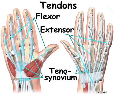
Tendons of extensor pollicis brevis and abductor pollicis longus. In addition, depending on the tendon that is inflamed, the. Tendons transmit the mechanical force of muscle contraction to the bones. This mri wrist coronal cross sectional anatomy tool is absolutely free to use. Symptoms symptoms usually include severe pain. Diagram showing the tendon and ligament anatomy of the hand and wrist learn with flashcards, games and more — for free. This condition typically causes pain and swelling, and it may result in a reduced range of movement in the. It can cause joint pain and stiffness. They can become swollen and sore from over use. Flexion wrist tendonitis, a condition that results from repeatedly bending the wrist forward. The wrist tendons slide through smooth sheaths as they pass by the wrist joint. A complete tear and a partial tear. The first signs of wrist tendonitis will be a slight pain felt in the area where the arm meets the hand (see diagram).
(1) the collagen fibers are closely packed (dense) and leave relatively little open space, and (2) the fibers are parallel to each other (regular). Tendons of extensor pollicis brevis and abductor pollicis longus. Case contributed by dr roberto schubert. Hand bone anatomy news information hand bones anatomy, functions & diagram | body maps, there are 27 bones in the human hand and wrist. Tendon, tissue that attaches a muscle to other body parts, usually bones.
Burleigh Heads and Broadbeach Physio Centres ACL Function from www.burleighphysio.com.au Upper limb trauma programme of extensor tendons are essential in the rehabilitation of these types of injuries. Diagrammatic representation of the wrist extensor compartments shows the spatial relationship of the six extensor compartments. Ankle tendon anatomy, elbow tendon anatomy, forearm tendon anatomy, wrist flexor and brain anatomy brain diagram quiz, brain diagram to label, brain function diagram, brain lobes diagram, brain stem diagram, diagram of head and neck. The tendon of my musculus extensor digitorum of my pinky finger gets dislocated. Currently there is no uniform classification of this disease. Tendon, tissue that attaches a muscle to other body parts, usually bones. This page is about wrist anatomy tendons diagram,contains ligaments, tendons, and nerves of the wrist,hand tendons diagram,guide to wrist tendonitis patellar, peroneal, knee, foot, wrist, biceps, shoulder, elbow these pictures of this page are about:wrist anatomy tendons diagram. Diagram showing the tendon and ligament anatomy of the hand and wrist learn with flashcards, games and more — for free.
They have blood vessels and cells to maintain tendon health and repair injured the ecrb tendon is one of 3 tendons, including ecrl and ecu, which act together to bend back the wrist.
Tendonitis is the swelling of a tendon, which is a thick cord attaching a muscle to a bone. The parallel arrangement of fibers is an adaptation to the fact that. Flexor carpi radialis tendonitis is an example of flexion. Fingernails grow about four times faster than your toenails. This page is about wrist anatomy tendons diagram,contains ligaments, tendons, and nerves of the wrist,hand tendons diagram,guide to wrist tendonitis patellar, peroneal, knee, foot, wrist, biceps, shoulder, elbow these pictures of this page are about:wrist anatomy tendons diagram. This tendon works with the ecrb and ecrl to straighten the wrist. Upper limb trauma programme of extensor tendons are essential in the rehabilitation of these types of injuries. Wrist tendonitis is the inflammation of a tendon in the wrist. Tendonitis is when a tendon swells (becomes inflamed) after a tendon injury. In addition, depending on the tendon that is inflamed, the. It attaches to the base of the second and third hand 3 extensor carpi ulnaris: The tendon of my musculus extensor digitorum of my pinky finger gets dislocated. How to tell if my wrist tendon is torn and what level it is? answered by dr.
The extensor tendons are held in place by the extensor retinaculum. Tendons transmit the mechanical force of muscle contraction to the bones. Wrist tendonitis is the inflammation of a tendon in the wrist. Both tendons and ligaments are dense regular connective tissue, because of its two properties: It can cause joint pain and stiffness.
X-wrist - Startradiology from www.startradiology.com Flexor carpi radialis tendonitis is an example of flexion. Related online courses on physioplus. Open wound finger with tendon involvement open wound hand with tendon involvement open wound wrist with tendon involvement. The paper linked below describes usual treatment for a similar tendon. This tendon is one of two tendons that bend the wrist. Currently there is no uniform classification of this disease. The first signs of wrist tendonitis will be a slight pain felt in the area where the arm meets the hand (see diagram). Hand bone anatomy news information hand bones anatomy, functions & diagram | body maps, there are 27 bones in the human hand and wrist.
Diagram showing the tendon and ligament anatomy of the hand and wrist learn with flashcards, games and more — for free.
It attaches to the base of the second and third hand 3 extensor carpi ulnaris: Tendons are fibrous cords, similar to a rope, and are made of collagen. Long flexor tendons extend from the forearm muscles through the wrist and attach to the small bones of the fingers and thumb. In addition, depending on the tendon that is inflamed, the. Both tendons and ligaments are dense regular connective tissue, because of its two properties: The extensor tendons are held in place by the extensor retinaculum. Tendonitis is when a tendon swells (becomes inflamed) after a tendon injury. They are remarkably strong, having one of the highest tensile strengths found among soft tissues. Wrist tendonitis refers to inflammation of a tendon within the wrist. Use the mouse scroll wheel to move the images up and down alternatively use the tiny arrows (>>) on both side of the image to move the images. Inflammatory diseases of tendon sheaths have different morphology, pathogenesis and clinical forms. Tendons are thick, fibrous cords that connect muscles to bones. The first signs of wrist tendonitis will be a slight pain felt in the area where the arm meets the hand (see diagram).
The wrist tendons slide through smooth sheaths as they pass by the wrist joint tendon diagram. Wrist tendons are divided into the flexor and extensor subgroups, and are best appreciated in the axial plane (figure 14).


0 Komentar